Karen Titus
March 2019—The standard riff for talking about a promising new cancer test should be familiar to anyone within sneezing distance of a laboratory: There’s no one-size-fits-all assay.
But if any test were to come close, it would be liquid biopsy.
Are clinicians eager to use it? Check.
Is it relatively simple to do (check) with fairly quick turnaround times (check)?
Does it work for solid and hematological tumors? Check and check. Across multiple specimen types—serum, urine, vitreous fluid, cerebrospinal fluid, stool? Quite likely.
Can it be used to characterize patients’ molecular profiles, monitor therapy, assess tumor evolution, identify resistance mechanisms, and detect early disease and minimal/measurable residual disease? Half a dozen checks.
Even if liquid biopsy does fall short of a one-size-fits-all assay, it’s doing a reasonable impression of a Swiss Army knife (if not Sergeant Troy’s sword fantastic, for those of you who are Thomas Hardy fans). The assay is dense with meaning, its rise enticing and swift.

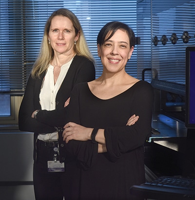
Dr. Maria Arcila (right) with Dr. Laetitia Borsu Valente, technical director for digital PCR and amplicon-based NGS assays, at Memorial Sloan Kettering Cancer Center. To date, the lung cancer group is the heaviest user of cfDNA testing in the clinical lab at MSKCC. (Photo courtesy of Jennifer Altman)
“It is amazing to see how rapidly this testing modality is being adopted,” says Maria Arcila, MD, director of the diagnostic molecular pathology laboratory at Memorial Sloan Kettering Cancer Center. Just a couple of years ago, she says, she did not foresee the current high demand for liquid biopsy testing from clinicians. But with the FDA’s approval of the first liquid biopsy test for EGFR in 2016, and newer and improved technology, applications are broadening, she says, “and one can only expect that the demand will keep growing.”
Liquid biopsy overcomes several of the shortcomings of tumor tissue biopsy. Aside from being readily accessible and a minimally invasive source of DNA, it addresses the problem of tumor heterogeneity, since circulating tumor DNA is a mixture of DNA derived from multiple sites and the tumor as a whole. “It is therefore potentially more representative of the malignant disease relative to a single localized biopsy,” Dr. Arcila says. Equally exciting is its use in longitudinal monitoring of patients for disease recurrence or for development of resistance mechanisms once a targeted therapy has been given. “You couldn’t do that before,” she says, without subjecting the patient to serial invasive biopsies and the associated risk.
It also addresses the drawn-out logistics of scheduling, performing, and processing a biopsy, which can stretch several weeks. With the faster turnaround time of a liquid biopsy, “you’re doing the patient a huge service,” Dr. Arcila says.
There are other benefits as well, and newer applications are being explored. “Circulating tumor DNA is dynamic and could be used to discover tumor changes and its evolution in real time,” she says. Positive molecular markers in ctDNA postoperatively, for example, could be used to assess for the presence of residual disease or metastases.
With liquid biopsy, that advantage joins another truth. Simply put, “Cancer is molecular,” said Mark Routbort, MD, PhD, professor of hematopathology, UT MD Anderson Cancer Center, in a presentation at the Association for Molecular Pathology annual meeting last November.
He’s been doing molecular sign-outs only since 2012, he told his audience, entering the field at the time when next-generation and massive parallel sequencing were emerging as a way to detect the activating locations, sequence variants, and copy number changes that are ubiquitous in human cancer—and not only in tissue. Around the same time came revived interest in the fact (first described by Mandel and Metais in 1948, he noted) that nucleic acids of normal and tumor cells circulate, and that it’s possible to detect elevated DNA in the serum of cancer patients. Echoing a point Dr. Arcila made in her own AMP presentation, Dr. Routbort said that sufficient DNA in a patient’s circulation most likely points to cancer.

Dr. Routbort
Little wonder that researchers have moved briskly to bring this knowledge to bear on clinical applications.
At Memorial Sloan Kettering, Dr. Arcila and colleagues have been using digital PCR to detect key genetic alterations in single genes such as EGFR and the resistance mutation T790M, as well as mutations in BRAF, IDH1, and IDH2.
Digital PCR, or dPCR, is a great technology, she says, for periodic monitoring of well-known genetic alterations. While many methods are available for assessing ctDNA, Dr. Arcila says dPCR and BEAMing have shown superior sensitivity. Based on published literature, dPCR sensitivity may be 0.1 to 0.01 percent. It’s also quite rapid and is an inexpensive technology, with no need for the specialized bioinformatics support that next-generation sequencing technology would require.
Sensitivity is based on the amount of DNA that can be recovered from a plasma sample, says Dr. Arcila, adding that the amount of cfDNA shed by a tumor varies from patient to patient. “That’s an important concept.” The rate of shedding of tumor DNA into the circulation depends on many factors, including the location, vascularity, and size of the tumor and the amount of necrosis, among others. “The more advanced the disease and the more metastatic sites the patient has, the more ctDNA they’re going to have,” Dr. Arcila says. (As a reminder, she adds, cell-free DNA encompasses both normal and circulating tumor DNA.)
Digital PCR, she explained in her AMP talk, is a biotechnology refinement of conventional PCR, used to clonally amplify and directly quantify nucleic acids. The key difference between dPCR and other PCR techniques is partitioning. Rather than running a PCR reaction in an entire tube, the test is run as thousands to millions of independent PCR reactions in discrete microdroplet-based compartments. By dividing the sample into so many droplets, the likelihood of having more than one DNA molecule being amplified in an individual droplet is quite low. That makes the assay precise, quantitative, and highly reproducible. Dr. Arcila called it unparalleled precision.
Digital PCR is relatively straightforward, she says, though like any assay, it has limitations and there are pitfalls. “We note that changes in the extraction protocols, for example, can have a major impact on how the assay performs,” she says, as can changes in temperature or humidity. Digital PCR is also not high throughput. “It is the type of assay one would do for mutations that are known to be hotspot variants and with well-defined roles in patient management.”
Memorial Sloan Kettering launched its dPCR journey three years ago with a test for EGFR T790M for patients with non-small cell lung carcinoma. Cell-free DNA testing is used as a screening test at the time of clinical suspicion of secondary resistance to EGFR tyrosine kinase inhibitors; only if the results are negative is the patient scheduled for a biopsy.“The most important thing to keep in mind is that there are limitations to a liquid biopsy,” Dr. Arcila says. “Very low levels of ctDNA, together with low overall cfDNA recovery from a plasma sample, may lead to false-negative results.” This may be particularly evident in certain scenarios, she says, such as assessing for resistance mutations, which are often subclonal compared with the original sensitizing mutation. “So to me, any negative result with cell-free DNA should be taken as a false-negative until proven otherwise, and close follow-up or rebiopsy may be warranted.”
Liquid biopsy samples, as it turns out, are much more difficult and variable than tumor biopsies. With the latter, “one can physically assess tissue suitability and presence or absence of tumor before testing is performed,” Dr. Arcila says. “With cell-free DNA, who knows? You just have to extract it, and you won’t know how much ctDNA is in your cfDNA sample unless the result is positive.” Guidelines and standardized methods for cfDNA qualification, quantification, and testing are not yet defined as they are for other molecular methods.
To date, “our lung cancer group is the center’s heaviest user of cfDNA.” Current lung molecular testing guidelines permit use of cfDNA testing if a biopsy cannot be obtained, she says, but other cancers don’t yet have similar guidance. “All of the questions right now are related to how fast each individual group introduces it into their guidelines.” What has been moving along at a brisk walk will likely turn into a trot, if not a run, once there are standardized protocols and methods, she says. “But there’s still a lot of work to do.”
In addition to the rapid dPCR tests, “our group has also developed a broad panel for cell-free DNA testing by next-generation sequencing with duplex unique molecular indexing called MSK-ACCESS,” which will soon be used in the clinical laboratory. The assay covers selected exons of 129 genes, as well as select copy number alterations and structural variants. “At this time, many institutions are or will be developing their own cell-free DNA assays. I don’t see it going backwards from here,” Dr. Arcila says.
While options abound, including comprehensive approaches using large hybrid-capture panels and exome sequencing, most clinical laboratories are using more targeted approaches to allow high depth of coverage of the most clinically relevant regions, according to Dr. Arcila. With dPCR, it may be possible to multiplex up to 15 mutations, although in a clinical setting a multiplex of even five will be challenging. Sensitivity is excellent, as she noted, and it is cost-effective for rapid genotyping and serial monitoring for specific, critical mutations. It also provides absolute quantification of mutant to wild-type copies and has minimal instrumentation requirements.
Using NGS panels provides a more comprehensive approach to known and unknown alterations. But, Dr. Arcila cautions, this approach is more costly and has longer turnaround times and results are semiquantitative. It will also require a specialized infrastructure to run the assays, as well as a team of experts to process and analyze results, including strong bioinformatics support.
That would be the bailiwick of Dr. Routbort, who’s been diving into cfDNA and related bioinformatics at MD Anderson, where he is also medical director of laboratory informatics and director of computational and integrational pathology. For panel-based cfDNA assays, he said, there’s been strong initial clinical evidence that serial monitoring of tumor-specific DNA may be significantly superior to imaging for predicting outcome and relapse.With cfDNA, he noted, “What you see in circulation may represent a mixture of tumor genotypes in a heterogeneous or a clonally evolving neoplasm.
“Now, that can be either an advantage or a disadvantage,” he continued. The circulation compartment may reveal either the most aggressive genotype or a mixture of genotypes, “so you may see things circulating that aren’t visible in the particular tumor section” obtained by biopsy for sequencing.
Dr. Routbort then sounded a warning. “You ignore the compartment issues at your own peril,” he told his AMP audience. And then, to much laughter, he noted that while it was hardly Theranos’ only problem, the company’s downfall arguably started with compartment issues—using capillary bloodstick samples versus those from central venous circulation led to testing inaccuracies.
The assumption is that tumor compartment(s) equilibrate DNA with plasma in a reliable manner, he said. While they may indeed be highly correlated, that doesn’t translate into linearity. Individual patient biology may be the key to whether labs can reliably interpret a negative finding.
Plunging into the data, Dr. Routbort offered some compelling evidence of liquid biopsy’s clinical value.
One MD Anderson group, as part of a DNA assay validation for thyroid carcinoma, looked at patients with RET M918T mutated medullary thyroid carcinoma. Some of the patients known to have RET M918T-positive tumors were negative (“completely undetectable,” says Dr. Routbort) in the assay, and some had very high circulating variant allele frequencies, with a wide range in between. “It’s orders of magnitude,” he said.
Looking at outcomes, the researchers found that patients with positive detectable circulating RET M918T had poorer prognosis. “If it’s negative at the time they’re tested—and these were all patients with metastatic disease—then the prognosis is much better,” Dr. Routbort said.
 CAP TODAY Pathology/Laboratory Medicine/Laboratory Management
