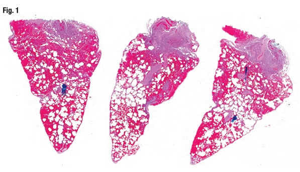
July 2019—Most fresh blood in lung wedge biopsies is artifact, but when it’s diffuse alveolar hemorrhage, the pathologist must call the clinician because DAH patients can go downhill fast. Maxwell L. Smith, MD, a consultant in the Department of Laboratory Medicine and Pathology at Mayo Clinic Arizona and associate professor, Mayo Clinic School of Medicine, shared that pearl from one of the 10 consultation cases he and Brandon T. Larsen, MD, PhD, co-presented in their CAP18 session on diagnosing interstitial lung disease. Their discussion of two of those cases follows.
Read More »Sidestepping pitfalls in diagnosing interstitial lung disease
June 2019—The pathologic approach to evaluating specimens as part of a workup for medical lung disease demands a different strategy than is typically used for the patient with a question of neoplasia, says Brandon T. Larsen, MD, PhD.
Read More »Peripheral blood evaluation: B12 deficiency, anemia, ET, CMML
April 2019—Diagnoses of essential thrombocythemia and chronic myelomonocytic leukemia were among those covered in four cases in a CAP18 session on practical challenges in peripheral blood evaluation.
Read More »Practical challenges in peripheral blood smear evaluation
March 2019—Unlike high-count monoclonal B-cell lymphocytosis, low-count MBL has a limited chance of progression to CLL and thus there is no need for follow-up. And a definitive diagnosis of T-cell large granulocytic leukemia requires demonstration of clonality.
Read More »Apocrine breast cancer, ESR1 mutations at center of tumor board review
February 2019—Two breast cases—one of apocrine carcinoma and androgen receptor overexpression and another of metastatic ER-positive cancer and an ESR1 mutation—were the focus of a molecular oncology tumor board session at CAP18.
Read More »Know them when you see them: parasites in tissue
February 2019—In one case, Dr. Ribes found herself intrigued by structures in a patient’s urine cytology specimen. A 36-year-old man from the Republic of Congo had developed dysuria, increased urinary frequency, and terminal hematuria. What they found in the Pap stain in cytology, she said, were large purple structures with the knob at the end.
Read More »Parasites in tissue: how to identify the structures
January 2019—Pathologists who aren’t microbiologists can provide a diagnosis of parasitic disease if they take into account parasite life cycles and tissue tropisms. Julie A. Ribes, MD, PhD, made that key point in cases she presented in her CAP18 session, “Update on Invasive Parasitic Infections for Surgical Pathologists.” Dr. Ribes added learning material to most of the cases, she said, but the cases come from parasites she has seen and known in her own professional life.
Read More »