 CAP TODAY Pathology/Laboratory Medicine/Laboratory Management

Karen Titus
July 2014—When it comes to molecular testing for acute myeloid leukemia, the approach seems more Montessori than military school.
There are some basic steps physicians should take, to be sure. Cytogenetics still shepherds patients into three prognostic groups: favorable, intermediate, and unfavorable. And several gene mutations—NPM1, CEBPA, FLT3, and KIT—alone or in combination, and with various cytogenetic associations, provide additional prognostic and therapeutic guidance.
But like the Cole Porter song, there’s a whiff of “anything goes” hovering about AML testing. No strict rules govern when testing—not just molecular—should be done, or where. When results do come in (each at its own pace), labs rely on their own creative solutions for coordinating the data. And in the face of seemingly endless molecular markers, physicians often pick their favorites, (lack of) evidence be damned.
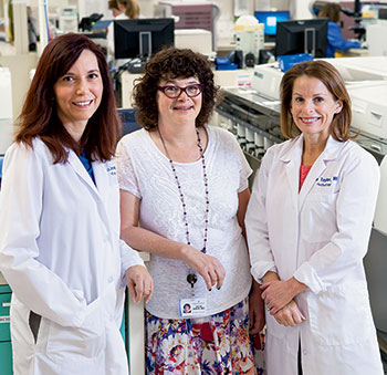
Dr. Melissa Cessna, left, with hematopathologist Ann Taylor, MD, right,
and oncologist Julie Asch, MD, associate director of the Intermountain Blood
and Marrow Transplant/Acute Leukemia Program. “Acute leukemias
are so diverse,” Dr. Cessna says, “and the testing seems to be exploding.”
“It’s hard for pathologists if every oncologist—and we have 30 or 40 here—wants something different,” says Rita Braziel, MD, professor, Department of Pathology, and director of the Hematopathology Division, Oregon Health & Science University, Portland.
Little wonder, then, that demand arose for a more formal guideline, addressing all acute leukemias; one is now being put together by an expert panel of CAP and American Society of Hematology members. The evidence-based guideline could be ready by early or mid-2015, says committee co-chair Daniel Arber, MD, the Ronald F. Dorfman, MBBch, FRCPath, professor of hematopathology, and vice chair of pathology for clinical services, Stanford University Medical Center.
But until then (and, frankly, even after), laboratories will have to continue their search for the best testing strategy. As the guideline committee and other pathologists know from experience, AML gives a decent imitation of perpetual motion. “It’s impossible to stop,” says Dr. Braziel. “Even if you take a standard subtype of AML, like acute promyelocytic leukemia [APL], which you think you know everything about—well, you don’t.”
During its literature search, the ASH/CAP guideline committee has discovered firsthand this unceasing aspect of AML, says Dr. Arber, who is also the medical director, anatomic pathology and clinical laboratory services, and co-associate director, molecular pathology laboratory. Recently Dr. Arber and his fellow co-chair, James Vardiman, MD, embarked on another round of data extraction from the articles (some 1,200 and counting). “That took quite awhile,” says Dr. Arber. “We didn’t have enough detail after the first round.” Another data refresh is likely at the end, he adds, acknowledging that newer data could likely support even stronger recommendations.
The initial push for the guideline came from Melissa Cessna, MD, a hematopathologist with Utah Pathology Services Inc. and Intermountain Healthcare, a community-based system. As Dr. Cessna observes, “Acute leukemias are so diverse, and the testing seems to be exploding. For community pathologists—and actually pathologists in any setting—understanding which tests should be done and how they’re used is important. But it’s hard to tease out.”
Dr. Cessna, who is on the committee, got her first taste of developing an AML testing algorithm when Intermountain expanded its bone marrow transplant service in 2006. The system now has a robust service for acute leukemia, she reports. Figuring out the right tests required working closely with the clinicians and balancing two competing but equally compelling demands: cost and choice. “I remember talking to one doctor who said, ‘I always like to have this marker,’” Dr. Cessna recalls. “She always had a ‘Did ya get?’ list—did you get this, did you get that?” Her reasons were sound. “You didn’t want to get stuck later not having a test that may have been relevant. But you were always guessing what might be relevant, and in that setting you would be shotgun ordering every test you might need.”
Intermountain’s strategy mimics air traffic by putting some tests in a holding pattern. Rather than ordering every FISH probe up front, Dr. Cessna and colleagues triage samples based on morphology and flow cytometry results. If they suspect APL AML, they then order stat FISH for t(15;17). Chromosome testing alone, she says, often provides much useful information. If the karyotype is complex, then the patient is known to have a poor prognosis, and additional FISH or molecular markers are unlikely to be relevant.

Dr. Arber
Molecular markers are “best proven only in certain situations,” Dr. Cessna continues. In patients with normal karyotype AML, FLT3, NPM1, and CEBPA will be prognostic. If FLT3 is positive, the prognosis will be poor. Mutated NPM1 indicates a good prognosis if there is no FLT3 mutation. Double-mutated CEBPA is associated with a good prognosis when FLT3 and NPM1 are negative.
Gone is the Old Country Buffet style of molecular testing in AML. Instead of laying out a spread, Intermountain now offers a series of tests, with each one requiring a clinical reason to move to the next “course.” “So for every new leukemia we just extract DNA and RNA, and there’s never a question of, ‘Did ya get this?’ Because we know we can add it on later,” says Dr. Cessna.
Doing so requires Dr. Cessna and colleagues to be in close contact with their reference laboratory as well as clinicians, however, and Dr. Cessna readily concedes Intermountain’s approach won’t work for everyone. Each lab, like Tolstoy’s unhappy families, is different.
Even in her own setting, Dr. Cessna sees the limitations. The hospital where she’s based serves a reference center, and it regularly sees patients who were previously diagnosed and perhaps treated with first-line induction therapy at another community hospital. Not all these patients have been tested for, say, FLT3, and they may not have had a DNA extraction. “We don’t have the capability to go back and add that, so you’re missing a piece of information that’s very important,” she says.
When Dr. Cessna and her colleagues created their algorithm, they soon identified that much of the acute leukemia testing was low volume. Even as a large community hospital, then, it would make sense to continue sending most of the molecular testing to an outside reference laboratory.
“We had a lot of discussion about what was practical,” Dr. Cessna recalls. “What was practical, and what we chose to employ, was to have our reference laboratory do DNA and RNA extractions.” In fact, they use two labs; one will hold material indefinitely, and the other for three weeks. “But we had to tell them what we needed, and they were willing to accommodate us.”
With AML specifically, she says, “We asked our reference laboratory, ‘What are the things that can be cryptic? What can be missed with a karyotype?’” In the resulting strategy, Dr. Cessna’s lab performs chromosome analysis, labeling it “high priority” so it can be triaged ahead of other cases. If the results are normal, it’s reflexed to three or four FISH probes known to alter prognosis, including inv(16), MLL gene rearrangement, and t(8;21). These results, in turn, will drive the molecular testing.
CEBPA is the most prognostic in the setting of a normal karyotype, negative FLT3, and negative NPM1 AML. “So if we have all those things, only then do we order the CEBPA,” Dr. Cessna says. KIT mutation testing is done only in cases with inv(16) or t(16;16) or t(8;21).
Having the lab hold material for possible later testing can slow turnaround times. Early on, Intermountain waited to order FLT3 until chromosome test results were available, but it now orders FLT3 at diagnosis, at the request of clinicians. Again, working closely with the reference laboratory paid off.
As for pushback from clinicians: “There was very little,” Dr. Cessna says. When the lab started looking into a new approach, it quickly became clear that many tests were being ordered unnecessarily, simply because oncologists were fearful of missing something. “But testing is expensive, and whether it’s relevant is dependent on what you see under the microscope,” Dr. Cessna says.
Intermountain offers so-called pathologist discretion for each type of test category. For flow cytometry, this is a “hold flow.” “We tell the lab whether or not to run it after we’ve reviewed the slides,” says Dr. Cessna. For cytogenetic studies, which include chromosome analysis and FISH, this will result in a “grow and hold,” where the laboratory will begin the cultures but not analyze them unless full analysis is requested. FISH can be added to a grow and hold or to a full chromosome analysis ordered up front within 90 days of collection. Grow and hold is a fraction the cost of the full study, she says.
“Adding FISH after you have the confirmatory diagnosis is also key,” Dr. Cessna says. “If you order based on what the diagnosis might be rather than what it is, there is a chance you’ll perform unnecessary or incorrect testing.” Take, for example, a patient who has pancytopenia, with AML in the differential diagnosis. If the AML panel is ordered up front, before the morphology has been reviewed, but the patient is later found to have metastatic breast cancer, then neither chromosome analysis nor FISH is needed. Ditto, she says, for flow cytometry—it would not be needed in this type of case, and it’s a very expensive test. “But if you order grow and hold, we can either not perform testing or add it after we’ve reviewed the slides. For molecular studies, we do a DNA and RNA extraction—the laboratory extracts the nucleic acids and stores them for us at -80°C.” She calls this approach “extract and store.”
In summary, she says, pathologist discretion results in (by category):