 CAP TODAY Pathology/Laboratory Medicine/Laboratory Management

Ann Griswold, PhD
October 2013—Culturing organisms is not always easy. Some microbes are fastidious. Others are below the limits of detection. And still others are just difficult to culture, for no discernible reason.
But set aside baffling cultures of the past because clinical microbiology is about to get more interesting than ever before.
“Clinical microbiologists should be aware that their laboratories are going to be increasingly asked to culture samples they’re not used to working with, samples that are not coming from patients,” says Matthew Arduino, MS, DrPH, FSHEA, lead microbiologist and chief of the Clinical and Environmental Microbiology Branch of the Division of Healthcare Quality Promotion at the Centers for Disease Control and Prevention’s National Center for Emerging and Zoonotic Infectious Diseases.
“They’re being asked to do more and more environmental microbiology.”
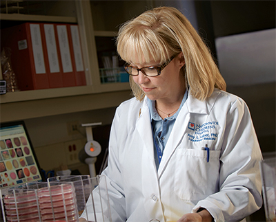
Dr. Amy Leber: “There’s a movement toward evidence-based medicine to verify the utility of our practices in the laboratory,” she says. “But in a lot of instances, there is no evidence.” [Photo: Dale Dong]
Whatever it is, Dr. Arduino says, the key to success lies in a careful approach. Here are the lessons learned by five clinical laboratorians who have faced these challenges head on.
Clinical laboratories have always played a critical role in patient safety. But focusing on patient safety no longer means focusing on specimens obtained directly from patients, says Alice Weissfeld, PhD, D(ABMM), president, CEO, and laboratory director of Microbiology Specialists Inc. (MSI), Houston. “We’re going to have to stretch and examine things that are not natural fits for the clinical laboratory to be able to prevent infections and really help the patients.”As a long-standing American Society for Microbiology delegate to the United States Pharmacopeial Convention, Dr. Weissfeld is leading the push for mandatory environmental testing of hospital compounding pharmacies. Frequent recalls of sterile products have shed light on this important issue. “Monitoring the pharmacy environment is as important as any patient culture,” she says. “We cannot let this fall by the wayside.”
Contamination in the pharmacy is a bigger problem than most people think, she notes. An article by Dr. Weissfeld and colleagues in the October issue of the Journal of Clinical Microbiology (2013;51[10]:3172–3175) examines five years of environmental sampling data from 30 hospital pharmacies and two freestanding pharmacies, and found that, on average, 35 percent of the pharmacies failed to meet USP standards every year. “When I ask pharmacists that I know how much contamination they actually think there is in the pharmacy, some say, ‘Oh, probably about one and a half percent.’ But that’s certainly not what we found.”
Dr. Weissfeld cites the recent outbreak of fungal meningitis that was traced to contaminated epidural steroid injections produced at the private NECC compounding pharmacy in Massachusetts. “Even though it was a freestanding pharmacy, some of that medication was administered in hospitals. We need to be prepared to help limit the damage from this type of problem.”
The USP has established recommendations for monitoring the pharmacy environment. “Under USP chapter 797, there are five activities that a clinical microbiologist can participate in with the pharmacy,” she notes. One is media-fill testing, which mimics the process of making sterile compounded preparations but uses tryptic soy broth (TSB) instead of the compound. The broth is then incubated for 14 days to check for contamination. Other activities include gloved fingertip testing, direct impaction air sampling, and surface sampling of hoods and compounding surfaces.
Dr. Weissfeld’s company, MSI, performs this work on a contract basis for hospitals. But to be most effective, she says, these activities should be performed on site by the hospital’s own laboratories, reserving the use of private labs for bioremediation and consulting. Most hospitals are open 24/7, and the test results would be available more rapidly from a hospital lab than from an environmental laboratory, which may only be open Monday through Friday, from nine to five.
“There’s a perception amongst the community that doing environmental sampling will somehow interfere with work on clinical cultures,” Dr. Weissfeld notes. “But this is not rocket science. These activities can be easily added. Yes, the media is slightly different [for environmental testing], but it’s not media that people who are clinical microbiologists would be unable to read.”
Private laboratories could continue to handle the challenging aspects—interpreting and addressing test results that exceed the limits described in the USP chapter. “I’m not asking clinical microbiologists to become bioremediators,” she says. “But I think the daily, weekly, and monthly sampling should be done in-house.”
Under increasing pressure to do more with less, hospital laboratories may find it difficult to add environmental testing to their plates. Environmental testing often falls into the category of things that do not have a direct impact on patient care, particularly in clinical laboratories that are dealing with cuts in staff, hiring freezes, and other constraints. “But when you’re looking down a list of things that you need to do, pharmacy testing is not one that should be thrown away,” she cautions. “Because if you are injecting a nonsterile product, the patient is either going to die or get very sick.”

Dr. Weissfeld
Dr. Weissfeld eschews the idea that pharmacists are in denial about the widespread problems with environmental contamination. “The disconnect is that they are out there by themselves. As with anything, this should be a team approach.”
One problem, she notes, is that most pharmacists have only a basic microbiology background and are not aware of the implications of pathogens for certain patient populations. That’s where close collaboration with a clinical laboratory would come in. “For example, if a hospital sees a lot of cystic fibrosis patients, the presence of Burkholderia spp may carry implications far beyond that of a gram-negative rod and source of endotoxin. In fact, infection with Burkholderia spp can have a profound effect on survival both pre- and post-lung transplantation. Clinical microbiologists know their patients and therefore would be able to respond quickly, if necessary.”
Performing environmental testing in-house could circumvent this problem by harnessing the clinical laboratory’s knowledge of its own patient population, and of the hospital’s resident pathogens. “It makes for a much more dynamic situation than if the pharmacy or an outside lab just counts numbers of colonies and identifies things, with no idea what any of it means. Microbiologists know what the results mean and can help the pharmacy interpret them.”
The next time Dr. Weissfeld surveys environmental sampling data from hospital pharmacies, she hopes to see a much lower failure rate. And that means finding better ways to support the hospital pharmacy. Organizations like the CDC, she says, are considering bringing together groups of clinical microbiologists and other stakeholders to establish guidelines for environmental testing in compounding pharmacies. Other efforts could be natural outgrowths of existing antibiotic stewardship programs, for example.
“If we can show pharmacists what they need to do—if our hospitals can better support them—then we can better protect patients as well as the hospital itself.”
Culturing unusual objects is simply part of the job for the Clinical and Environmental Microbiology Branch of the CDC. Microbiologists in the branch perform outbreak support, environmental surveillance, preparedness research, and susceptibility testing of resistant organisms such as carbapenem-resistant Enterobacteriaceae and methicillin-resistant Staphylococcus aureus.“If it’s epidemiologically linked, we will sit here and figure out how to do it,” Dr. Arduino says. Though he has faced many daunting tasks, one of the most memorable involved a large outbreak in a neonatal ward.

Dr. Arduino
The outbreak was traced back to instruments used to humidify air for the infants’ CPAP machines. When one of the suspect devices was shipped to the CDC for inspection, Dr. Arduino and his colleagues discovered that Ralstonia pickettii—a water-borne organism with a penchant for nutrient-poor environments—had formed a biofilm on the inside of the device. Sure enough, cultures from the instrument matched those from the infants. But how the pathogen made its way from the device to the patients’ airway remained a mystery. “There was a membrane, and we didn’t know how [the pathogen] was crossing through to the patient. It should have been a separate circuit,” Dr. Arduino recalls. Finally, the team unearthed an important clue: “We found out that the company manufactured this device overseas, and that one of the final steps before they shipped it was rinsing it with tap water.”
From this and countless other experiences, Dr. Arduino has learned an important lesson about human nature: “You can invent a better mousetrap but people will always find a way to circumvent it,” he says. Most problems occur when workers in manufacturing and clinical settings eschew microbiologists’ appreciation for cleanliness. He has seen a number of examples over the course of his career.
On Dr. Arduino’s first day of work at the CDC, he investigated an outbreak linked to a dialysis facility in California. Multiple patients had developed systemic infections with abscesses.
“When I first began investigating dialysis outbreaks, more than half of the facilities were performing manual reuse on the artificial kidneys,” he recalls. “There was somebody in a back room, usually a low-paid technician, who was rinsing the residual blood from the dialyzer, doing a pressure test, and then filling it with germicide so that it could be used on that same person again when they came in for the next session.”
The investigation revealed that the dialysis water was contaminated with a mycobacterium (Mycobacterium abscessus). When the CDC team dug further, they learned that the facility had recently switched from formaldehyde as its germicide to peracetic acid—but they were only using half the required strength. “The salesperson had said, ‘You know, this stuff is really good. The label says to use this concentration, but you can use half,’” he recalls. But that wasn’t the only precipitating factor. Dr. Arduino and his colleagues observed the facility’s cleaning process and realized that the technicians were filling the dialyzers with only half the recommended amount of germicide. The residual water remaining in the dialyzer diluted the germicide further, and the water used to rinse the dialyzers and prepare the disinfectant contained mycobacteria, which were not killed by the disinfectant placed into the dialyzer. The resulting outbreak was another byproduct of misinformation and inadequate training.
Communication between all workers in the clinical environment, including janitorial staff, is crucial, he says. In another outbreak he investigated—this one unrelated to dialysis—the only common exposure was a portable x-ray machine. The CDC obtained a sponge sample from various parts of the machine and recovered Acinetobacter baumannii from the surfaces that touched the patient.
“And then, the question was asked, ‘Who cleans the machine?’ Nursing said, ‘We don’t clean. That’s housekeeping.’ Housekeeping said, ‘No, no. That’s an instrument. We don’t touch those. That’s nursing.’ So the machine was never disinfected between uses,” he says.
A career’s worth of experiences like this one have taught Dr. Arduino a few things about culturing challenging objects. Most importantly, he says, a negative result doesn’t always mean the culture is negative. It’s essential to use techniques and media appropriate to the object being cultured, and to realize when an outdated or inappropriate technique might be raising the limit of detection.
As evidence, he points to the gradual evolution of standards for testing the water used in hemodialysis. History has shown that the ability to detect contaminants depends in large part on the methods and reagents used—and on the willingness to break from tradition in favor of techniques better suited to a novel task.
Today, most dialysis samples are sent to an environmental laboratory or a dialysis company laboratory for testing. But when clinical microbiology laboratories were first faced with the challenge of testing dialysis fluids in the early 1980s, Dr. Arduino says, standard clinical culture methods hindered the detection of dialysate contaminants, leading to many false-negatives.
In one outbreak, Dr. Arduino recalls, several dialysis patients developed fever, chills, and clear signs of bacteremia despite negative dialysate cultures. It turned out that the samples were cultured on blood agar, the standard substrate for patient specimens. “But microbes that you find in water or dialysate tend to be nutrient-poor,” Dr. Arduino says. “So when they inoculated a chocolate plate or a blood agar plate, the organisms would actually die because of the richness of the media.”
In addition to the rich agar, many clinical microbiologists at the time were using calibrated loops, which are typically used for urine analysis, to plate the dialysate samples. As a result, they missed the cutoffs for detection, Dr. Arduino says.
Because of this long and complicated history, he worries that some pathogens will continue to evade detection by routine screening due to inconsistencies in the practices various labs use. “Some laboratories are still doing membrane filtration; some are doing spread plates,” he notes.